Research
Neural mechanisms of spatial orientation
Our ability to keep a stable perception of the world is essential for nearly everything we do. This ‘orientation constancy’ allows us to stay balanced and make accurate movements. Spatial orientation refers to our awareness of where the body is in relation to the environment. Even though our eyes, head, and body are constantly moving, we naturally maintain a steady sense of body orientation correctly aligned with the world. When this system fails, it often n leads to dizziness, disorientation, and loss of balance.
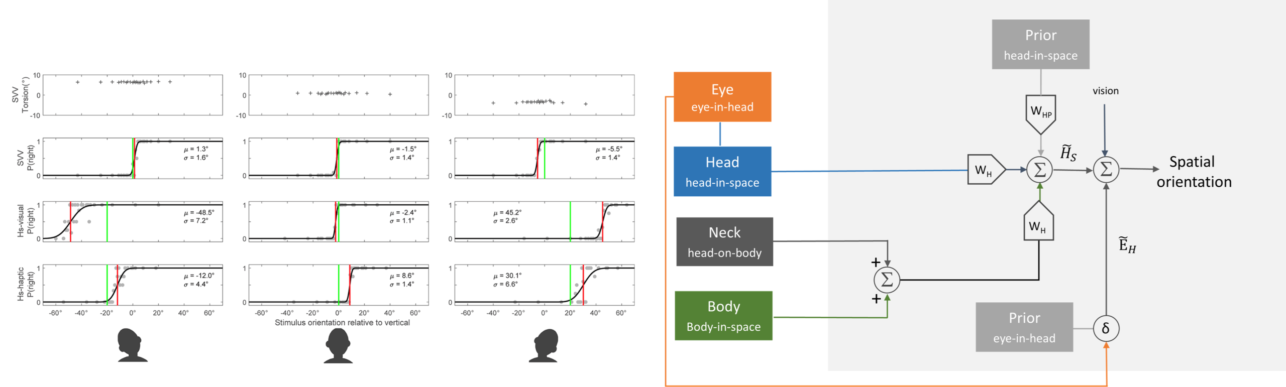
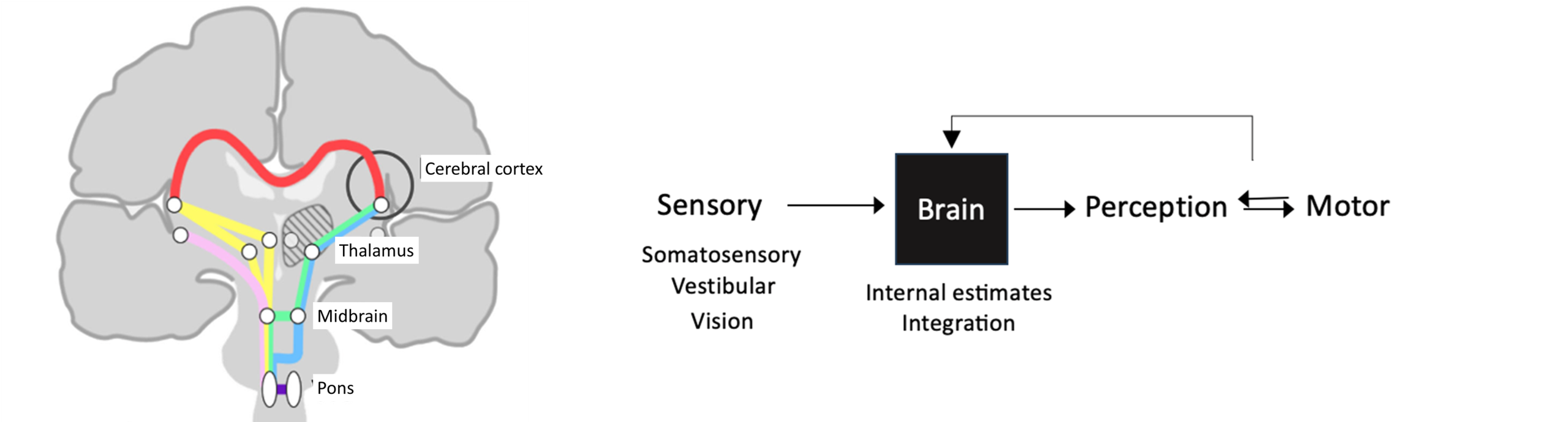
Our stable perception of spatial orientation is made possible because the brain integrates information from vision, the vestibular system, and proprioception to construct the orientation of the body within the external environment. To achieve this, the brain must solve a difficult problem: each sensory signal is encoded in different frame of reference. For instance, even with a simple head tilt, the reference frames of the eyes, head, and visual world become misaligned, yet our perception remains stable relative to gravity. This means the brain –much like an optimally tuned sensorimotor system– creates a unified internal reference frame to keep our experience of the world steady. In our lab, we study the neural mechanisms that support these key aspects of spatial orientation. We combine psychophysical experiments with computational models that examines how the brain transforms sensory signals into coherent perceptual orientation of the surrounding environment.
Eye movement control and interaction with balance and spatial orientation
Eye movements are represented throughout the brain, from the brainstem to the cerebral cortex. The brain controls many dynamic features of these movements, including their timing, velocity, accuracy, and trajectory. Because these processes are so tightly linked to neural function, clinicians and neuroscientists routinely use eye movements as a window into brain activity.
Why study eye movements?
- Functions that are well understood and organized into recognizable subclasses, each with specific physiological properties and anatomical substrates linked to inner-ear functions.
- Provides useful biomarkers in neurological disorders, helping to establish diagnoses, gauge disease severity and progression, and monitor responses to treatment or rehabilitation.
- Easy to stimulate, measure, and quantify in experimental studies.
- Well suited to rigorous mathematical and computational modeling.
- Ideal for studying higher cognitive functions such as prediction, memory, attention, learning, adaptation, and decision-making.
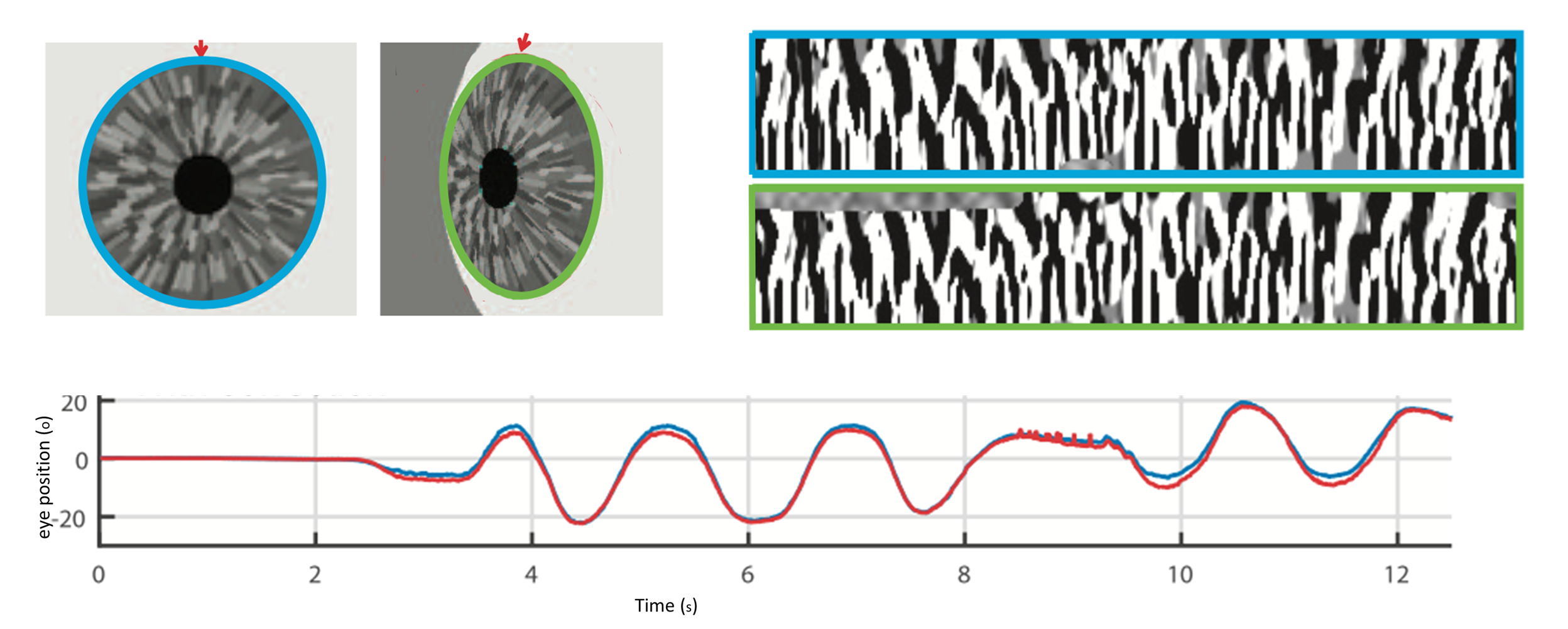
In our lab we use various techniques to measure eye movements with great precision, including dual magnetic scleral search coils and video-oculography. These tools allow us to track even very small or fast movements in a reliable and quantitative way. We also work on developing new recording and analysis methods to better support both research needs and clinical applications.
Functional mapping of neural networks for perception of spatial orientation
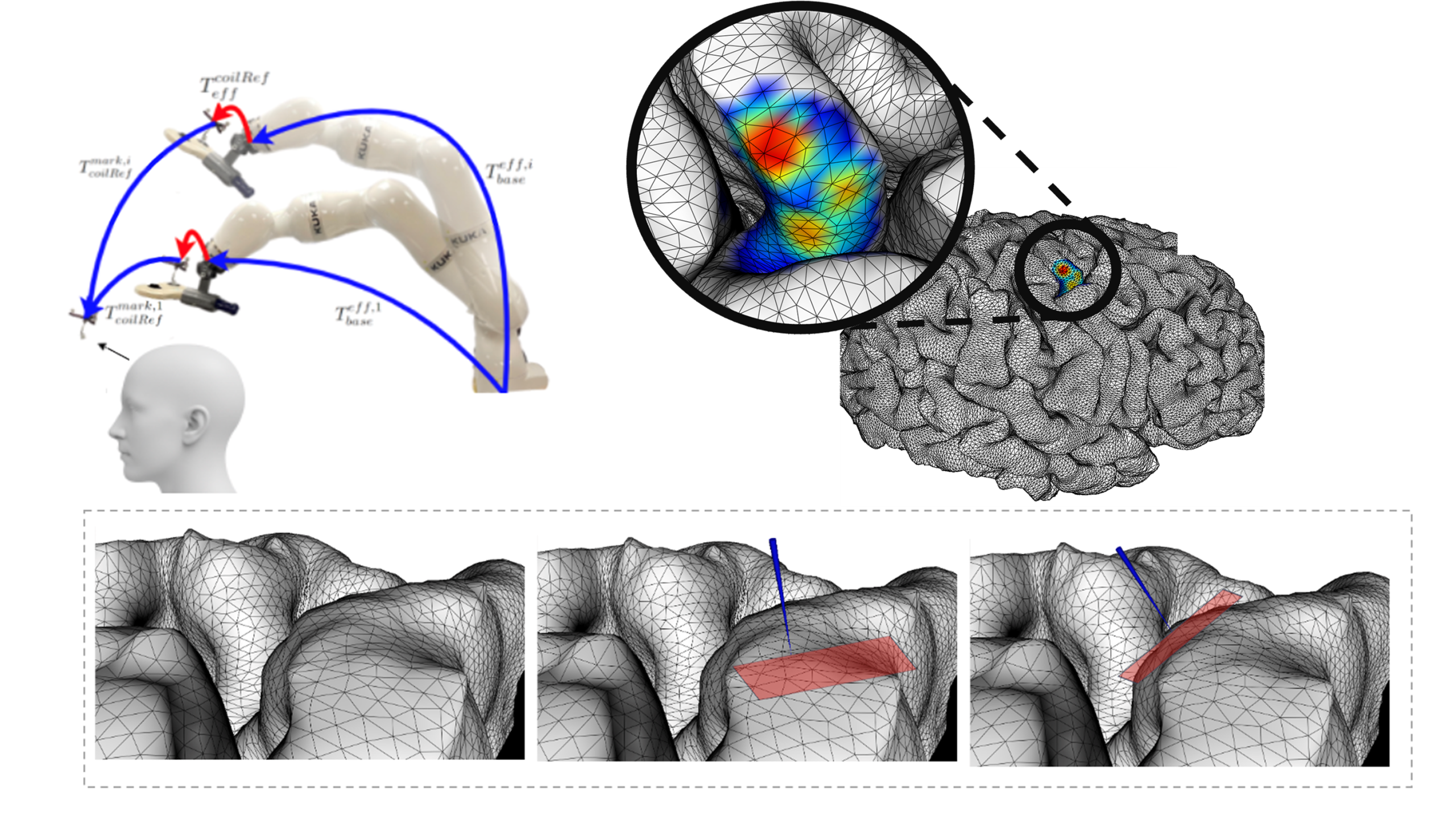
We study the complex neural processes involved in sensory processing and integration that give rise to our perception of spatial orientation. To investigate these mechanisms, we have developed a neuronavigation framework using detailed segmentation of brain geometry to probe specific brain areas. The system integrates two core components. First, a co-registration method aligns each participant’s head with their 3D MRI model at high spatial accuracy. Second, a refined planning algorithm that allows setting up a target position for transcranial magnetic stimulation (TMS) and real-time bran stimulation. Because this method depends on accurate modeling of cortical topography, it requires stable and precise TMS coil placement. Achieving such precision manually is challenging as small shifts of the head or TMS coil can alter the alignment of the magnetic field, introducing variability in stimulation accuracy and reducing efficacy. By integrating a robotic system, we overcome many of these limitations. The robot maintains highly stable and reproducible TMS coil placement, particularly during long TMS sessions. This precision is especially critical for topography-guided neuronavigation, where effective stimulation depends not only on selecting the correct anatomical target and coil orientation, but also on maintaining that orientation consistently throughout the entire stimulation session.
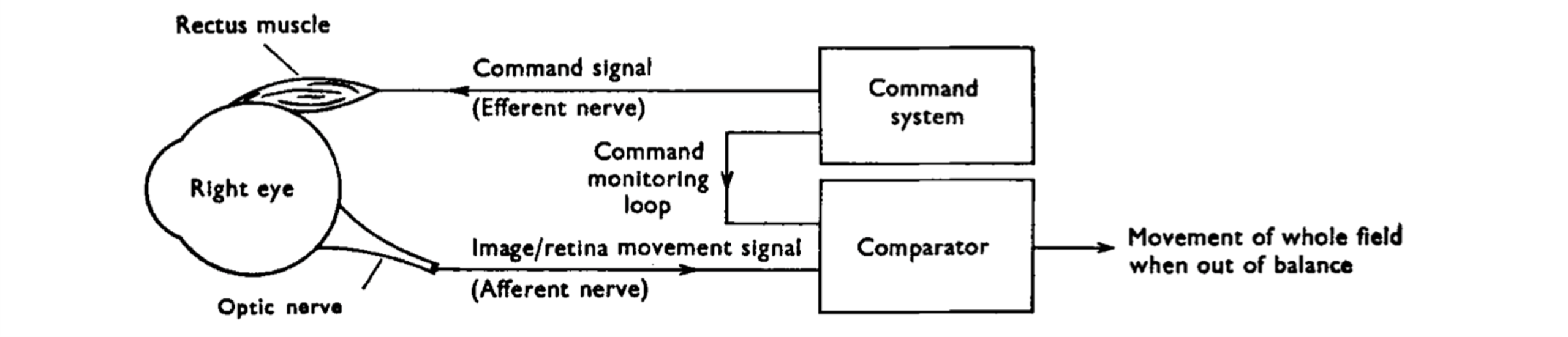
Neural mechanisms of visual stability
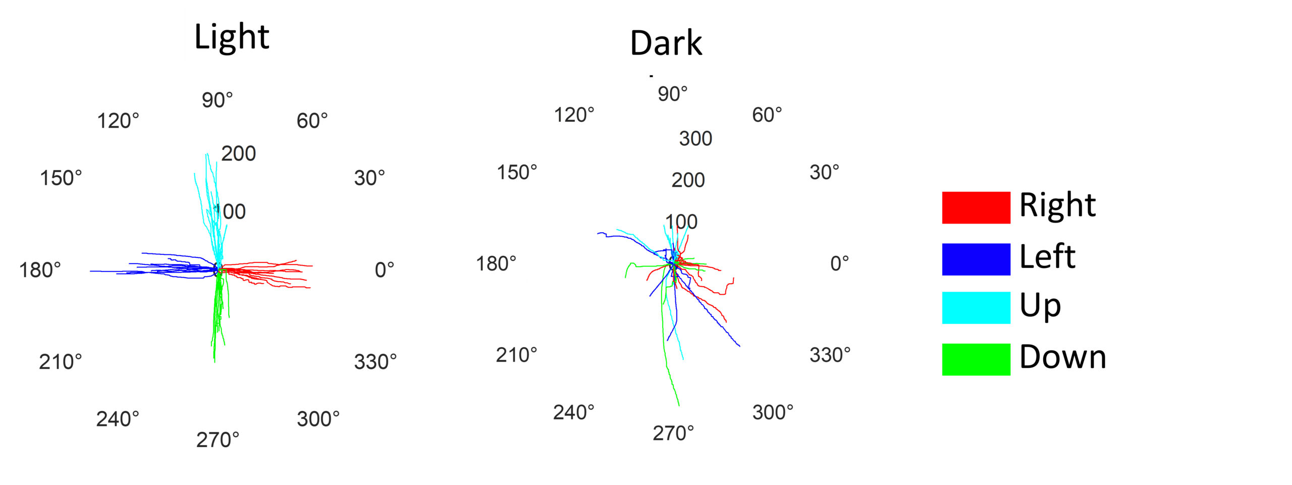
In natural viewing conditions, the brain can optimally integrate retinal and extraretinal signals to maintain a stable visual perception. These mechanisms, however, may fail in circumstances where extraction of a motion signal is less viable such as impoverished visual scenes. This can result in a phenomenon known as autokinesis in which one may experience apparent motion of a small visual stimulus in an otherwise completely dark environment. We investigate how autokinesis influences human perception of motion using novel methods in which observers manually report perceived motion. This work has important implications for understanding how the brain stabilizes visual representations and how eye movements, the resulting motion on the retina, and the brain’s processing of these signals collectively shape the accuracy of perceived motion.
Neural mechanisms of dizziness, imbalance, and spatial disorientation in disease sates
Dizziness is a common complaint across many clinical settings and remains a diagnostic challenge, even with modern advances in medical technology and imaging. Patients frequently describe misperceptions of self or environmental motion, abrupt feelings of disorientation, imbalance, or tilt. These complex symptoms, broadly characterized as dizziness or vertigo, often arise as the brain must continuously construct and update an accurate representation of the world by integrating different sensory cues. Our research examines how these integrative processes break down in disease states. We focus on key functions—such as motion perception, spatial orientation, visuospatial attention, and spatial awareness—that are essential for controlling body movement and maintaining orientation in the environment. Growing evidence from structural and functional brain studies suggests that these disorders reflect disruptions within distributed neural networks responsible for multisensory and sensorimotor integration, rather than isolated deficits in a single sensory pathway. By identifying the underlying mechanisms that give rise to symptoms in different disease states, we can develop more targeted treatments that directly address these specific neural dysfunctions.
Diagnostic tools for accurate clinical assessment of eye movement and vestibular functions
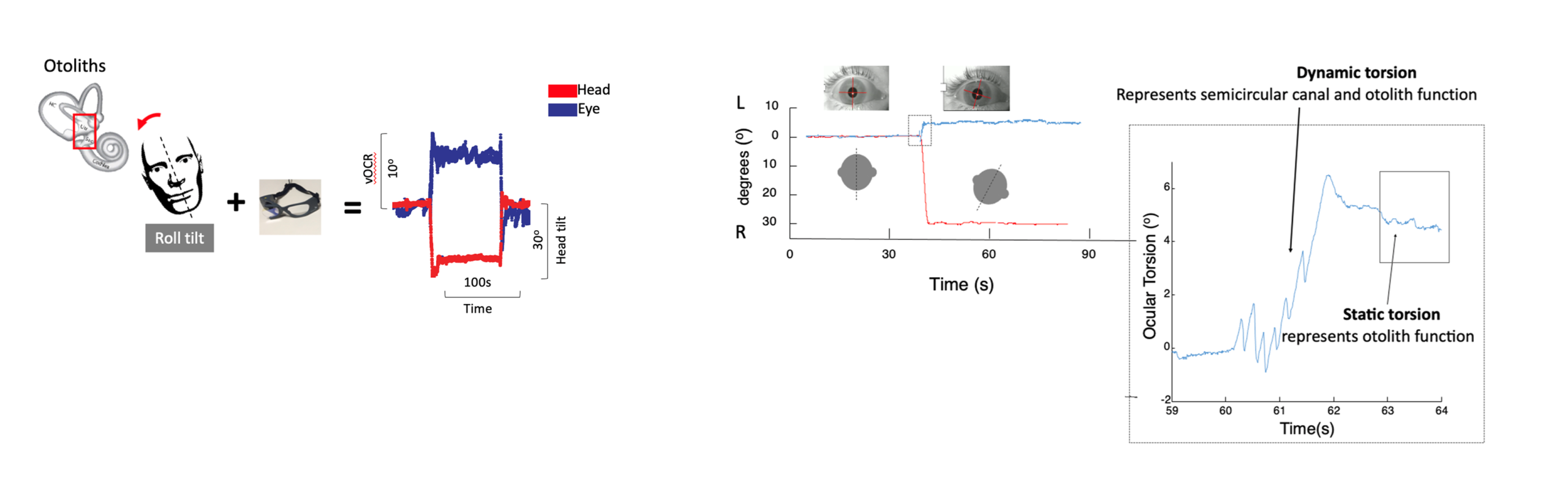
A normally functioning vestibular system is essential for spatial orientation, balance, and maintaining a stable gaze during head motion. Accurate clinical assessment of eye movements and vestibular function depends on specialized diagnostic tools that capture the dynamic interactions between the visual and balance systems. Recent advances in lightweight video-oculography (VOG) have transformed vestibular evaluation by enabling precise, quantitative measurements of eye movements at the bedside. Traditionally, vestibular assessment has focused primarily on semicircular canal function using tests such as caloric stimulation and rotational chair evaluation. The portability and improved resolution of modern VOG systems have expanded their integration into clinical practice, significantly enhancing diagnostic capabilities. Building on the underlying physiology of otolith–ocular pathways, we have developed a VOG-based test to assess otolith function clinically. This method, known as video ocular counter-roll (vOCR), measures the torsional vestibulo-ocular reflex during lateral head tilt and provides a practical, objective approach for evaluating otolith-ocular function.
Diagnostic tools for accurate clinical assessment of balance and posture
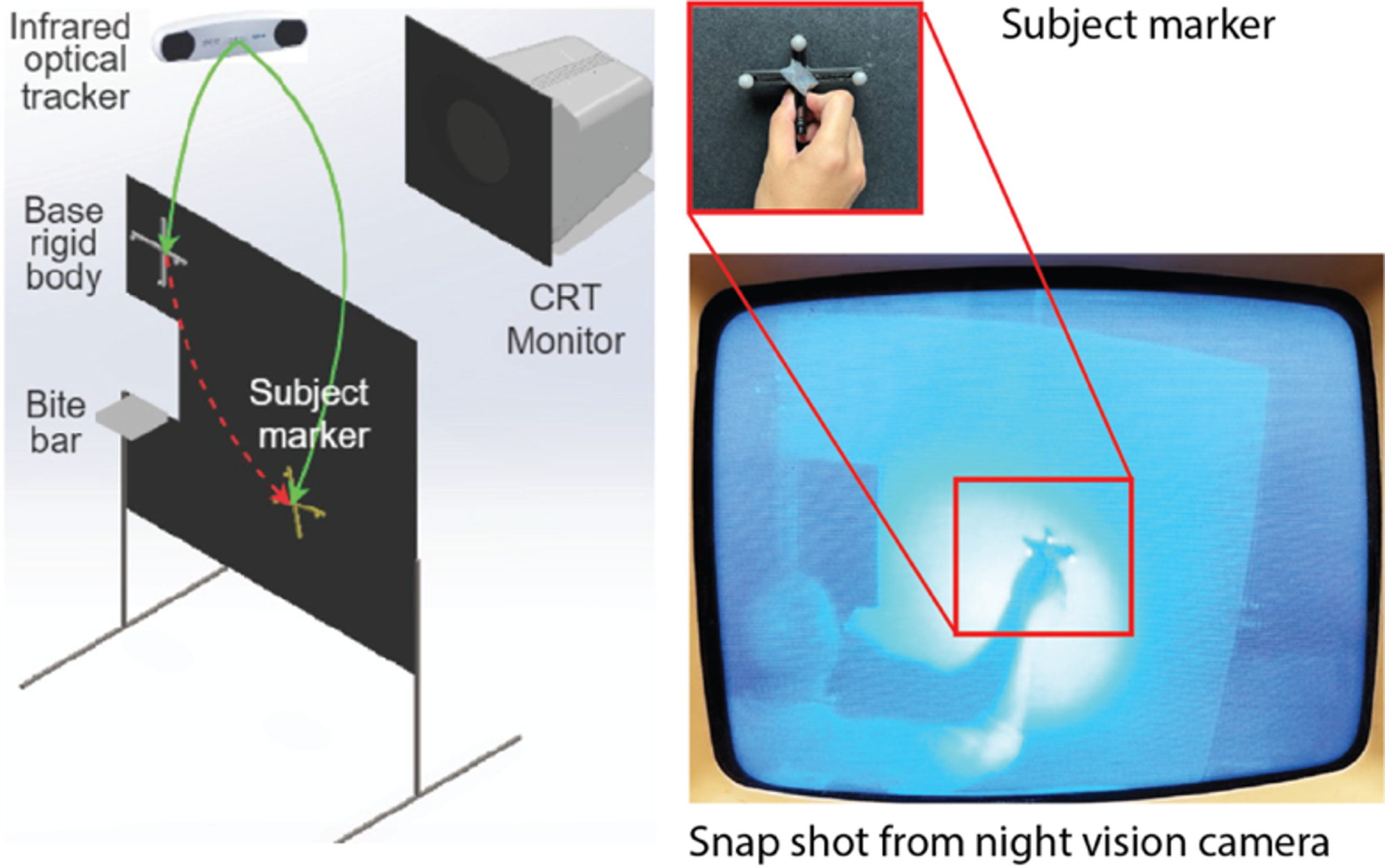
Gait and posture analysis are essential parts of evaluating balance, providing important information that cannot be captured by vestibular or eye-movement tests alone. By studying how a person maintains posture or walks, one can identify fall risk, recognize compensatory strategies, and gain insight into how the sensory and motor systems work together to keep a stable balance. We use different approaches including inertial measurement units (IMU) and video-based tools to study 3D body posture and motion. These methods can measure subtle changes in balance control and be integrated with other measurements to provide a comprehensive assessment of balance functions. We have developed a marker-less video system that uses a deep learning pose-estimation model to track body movements in three dimensions. This technology provides an accessible method for assessment of balance in both clinical and real-world.
